RESEARCH
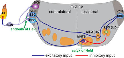
Auditory information is transmitted from the inner ear to the brain by spiral ganglion neurons (SGNs), which extend peripheral processes towards hair cells (HCs) in the cochlea and central processes through the eighth nerve and into the auditory brainstem. Upon entering the brainstem, the central process of each individual SGN bifurcates. The descending branch navigates through posteroventral cochlear nucleus (PVCN) into the dorsal cochlear nucleus (DCN) and makes standard bouton contacts with a variety of target neurons. In contrast, the ascending branch projects to the anterior ventral cochlear nucleus (AVCN) and elaborates an extraordinary large synaptic terminal, known as the endbulb of Held, on the bushy cell neuron (SBC/GBC) (please refer to the figure).
Development of the Endbulb of Held synapse
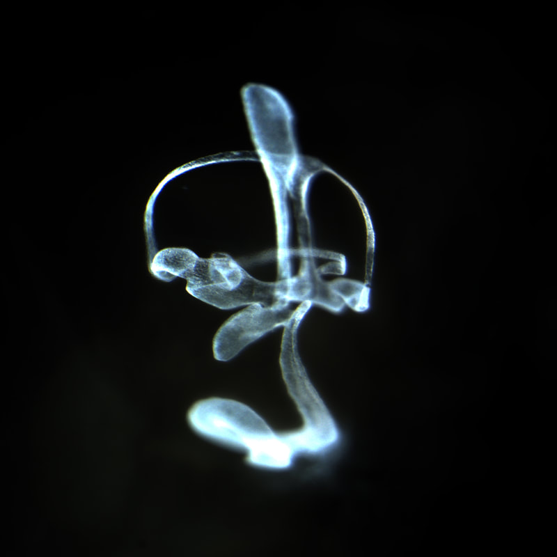
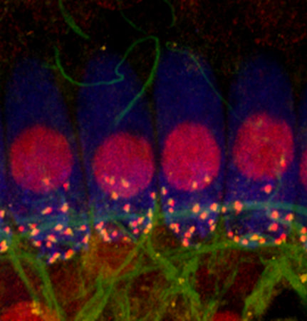
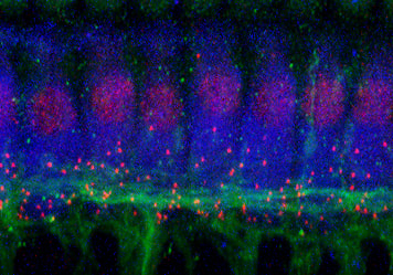
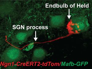
Auditory neural circuits use binaural cues to accurately locate a sound source in space and rely on exceptionally fast and precisely timed neurotransmission mediated by the specialized large endbulb synapses. However, little is known about how this giant synapse develops. We can use genetic approaches to sparsely label individual SGN central processes and the associated endbulb synaptic terminals (please refer to the figure). Using this strategy, we will reveal the cellular events of how and when the auditory circuit develops the Endbulb of Held, including outgrowth of SGN central processes, initial targeting of postsynaptic bushy cells in the VCN, the beginning of synaptic formation, and the maturation of the endbulb synapse.
Auditory neural circuits use binaural cues to accurately locate a sound source in space and rely on exceptionally fast and precisely timed neurotransmission mediated by the specialized large endbulb synapses. However, little is known about how this giant synapse develops. We can use genetic approaches to sparsely label individual SGN central processes and the associated endbulb synaptic terminals (please refer to the figure). Using this strategy, we will reveal the cellular events of how and when the auditory circuit develops the Endbulb of Held, including outgrowth of SGN central processes, initial targeting of postsynaptic bushy cells in the VCN, the beginning of synaptic formation, and the maturation of the endbulb synapse.
Cellular and molecular basis of tonotopic map formation
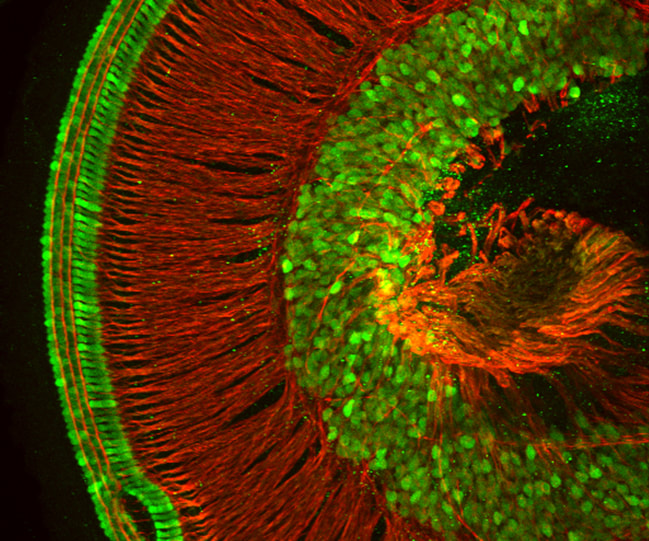
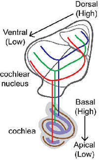
In the cochlea and cochlear nucleus, SGN cell bodies and their central fibers are arranged in a tonotopic gradient according to frequency responses. Neurons responding to high frequency sounds are located at the basal portion of the cochlea and project their central processes to dorsal regions of the cochlear nucleus, while neurons convey information of low frequency sounds are located at the cochlear apex and send their central projection to ventral portions of the cochlear nucleus (please refer to the figure). We can use genetic approaches to specifically label SGNs responding to different sound frequencies. The innervation patterns between high- and low-frequency SGN fibers will be compared at different stages to determine the cellular mechanisms of tonotopic map formation. We will also identify molecules that are involved in the tonotopic map formation.
In the cochlea and cochlear nucleus, SGN cell bodies and their central fibers are arranged in a tonotopic gradient according to frequency responses. Neurons responding to high frequency sounds are located at the basal portion of the cochlea and project their central processes to dorsal regions of the cochlear nucleus, while neurons convey information of low frequency sounds are located at the cochlear apex and send their central projection to ventral portions of the cochlear nucleus (please refer to the figure). We can use genetic approaches to specifically label SGNs responding to different sound frequencies. The innervation patterns between high- and low-frequency SGN fibers will be compared at different stages to determine the cellular mechanisms of tonotopic map formation. We will also identify molecules that are involved in the tonotopic map formation.
